
EEG vs fMRI: Privacy, Data Security and Neurotechnology Attack Surfaces
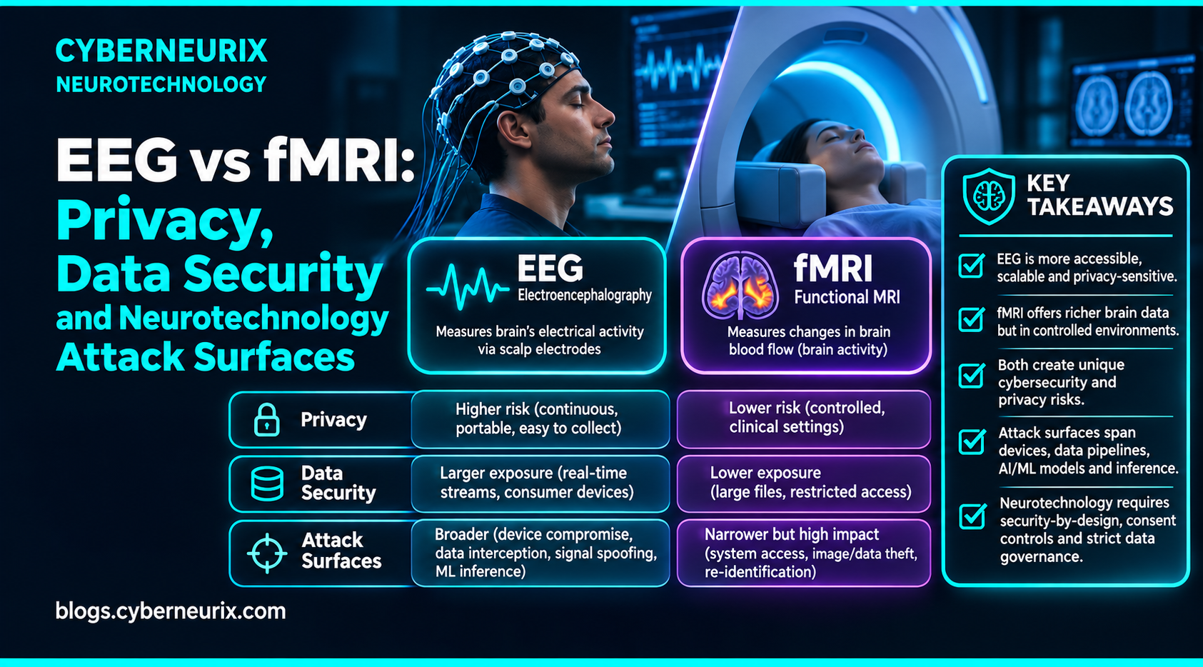
Key Takeaways
- EEG and fMRI represent opposite trade-offs in neuroimaging: EEG offers high temporal resolution (milliseconds) with low spatial fidelity, while fMRI provides high spatial resolution (millimeters) with slow hemodynamic lag.
- Security threat models must follow the acquisition architecture: EEG is vulnerable to wireless pairing hijacking, while fMRI is vulnerable to hospital PACS network compromise.
- Portable EEG generates continuous, longitudinal biophysical data streams; fMRI produces massive episodic volumetric scan files (NIfTI/DICOM).
- Metadata leakage in fMRI imaging headers regularly exposes patient clinical records and biometric facial reconstructions.
- Both modalities require strict cryptographic separation between raw neuroimaging files, derived feature spaces, and patient demographic identities.
The Dangerous Myth of Uniform 'Brain Data'
In legal policy briefs and data privacy debates, 'brain data' is frequently discussed as though it were a single, uniform technical commodity. Regulatory drafts attempt to craft blanket protection rules covering any digital record derived from the human nervous system. Yet in technical reality, there is as much difference between electroencephalography (EEG) and functional magnetic resonance imaging (fMRI) as there is between a real-time network packet capture and a 50-gigabyte database backup.
EEG and fMRI measure completely different physical phenomena. EEG captures rapid, microvolt-scale electrical potential differences directly generated by cortical pyramidal neurons at millisecond speed. In contrast, fMRI measures the blood-oxygen-level-dependent (BOLD) hemodynamic response—a slow, indirect metabolic echo of neural activity occurring over several seconds. Because their physical sensors, hardware enclosures, and data formats are radically different, their cybersecurity vulnerabilities and privacy threat models share almost nothing in common.
For security engineers, clinical IT architects, and privacy officers entering the neurotechnology domain, conflating these two modalities is a critical design error. Hardening neural interfaces requires tailoring defenses to the exact physical acquisition pipeline.
Deep Dive: Comparing the Technical Pipelines & Attack Surfaces of EEG and fMRI
1. Signal Physics & Pipeline Mechanics
A 2026 technical review published in the Journal of Neurophysiology establishes the structural dichotomy between the two modalities:
- Scalp Electroencephalography (EEG):
- Physical Phenomenon: Direct measurement of post-synaptic ionic currents across large cortical assemblies.
- Resolution: High temporal resolution (<10 ms), low spatial resolution (~several centimeters, heavily distorted by skull bone volume conduction).
- Data Velocity: Continuous, low-bandwidth real-time time-series streams (256–1000 samples/sec per channel).
- Functional Magnetic Resonance Imaging (fMRI):
- Physical Phenomenon: Radiofrequency excitation of hydrogen nuclei within a strong magnetic field (1.5T to 7T), measuring magnetic susceptibility shifts between oxyhemoglobin and deoxyhemoglobin (the BOLD response).
- Resolution: High spatial resolution (1–2 mm 3D isotropic voxels), poor temporal resolution (1–3 second repetition time [TR] reflecting hemodynamic lag).
- Data Velocity: Massive, episodic volumetric image files (often 500 MB to several GBs per clinical scan session).
fMRI anatomical scout scans (T1-weighted structural MRI) capture detailed facial tissue contours. An attacker with access to a 'de-identified' neuroimaging file can run standard 3D surface rendering software to reconstruct the subject's exact facial likeness, instantly matching it against public photo databases.
2. The EEG Attack Surface: Mobile, Wireless & Continuous
Portable EEG systems—whether clinical ambulatory monitors or consumer focus headbands—exhibit attack surfaces typical of IoT and edge devices:
- Wireless Transport Interception: Unencrypted Bluetooth or proprietary 2.4 GHz RF links allow proximate eavesdroppers to sniff raw brainwave telemetry.
- Pairing & Authentication Abuse: Lack of cryptographic mutual authentication enables malicious devices to inject spoofed sensor packets into decoding apps.
- Mobile SDK & Application Vulnerabilities: Third-party neuro-app SDKs embedded in consumer smartphones often exfiltrate background brainwave features to commercial advertising trackers.
- Longitudinal Cognitive Tracking: Continuous daily wear creates rich behavioral profiles detailing sleep quality, focus states, and emotional reactivity over months.
3. The fMRI Attack Surface: Clinical PACS, DICOM & Workstation Compromise
fMRI systems operate inside high-security hospital imaging suites, presenting enterprise clinical vulnerabilities:
- Scanner Console Workstations: MRI scanner control consoles frequently run legacy, unpatched operating systems (Windows 7/10 embedded) to preserve regulatory medical clearance.
- DICOM & PACS Infrastructure: Neuroimaging scans traverse clinical hospital networks via the Digital Imaging and Communications in Medicine (DICOM) protocol, historically lacking native encryption or zero-trust access control.
- Research Data Repositories & NIfTI Headers: Neuroscientists convert clinical DICOM files to NIfTI (.nii) format for processing. Research pipelines frequently overlook embedded DICOM header metadata containing patient names, medical record numbers (MRNs), and exact scan dates.
- Reconstructive Mental Privacy: Recent breakthroughs in deep learning and diffusion models allow researchers to reconstruct approximate visual scenes and semantic concepts directly from high-resolution visual cortex fMRI voxels.
| Dimension | Electroencephalography (EEG) | Functional Magnetic Resonance Imaging (fMRI) |
|---|---|---|
| Physical Signal | Post-synaptic ionic electrical currents | Hemodynamic blood-oxygen-level changes (BOLD) |
| Temporal Resolution | Ultra-high (<10 milliseconds) | Slow (1–3 second hemodynamic response lag) |
| Spatial Resolution | Low (~cm, subject to skull diffusion) | Ultra-high (1–2 mm 3D isotropic voxels) |
| Hardware Footprint | Lightweight, battery-powered wearable headset | Multi-ton cryogenic superconducting magnet suite |
| Primary Transport | Wireless RF (Bluetooth Low Energy, Wi-Fi) | Gigabit Ethernet, optical DICOM hospital fiber |
| Core Attack Surface | BLE sniffing, mobile app SDKs, rogue pairing | PACS servers, DICOM metadata, scanner OS exploits |
| Biometric Leakage | Cognitive workload, focus state, P300 identity | Facial 3D surface reconstruction from anatomical voxels |
CyberNeurix Unique Angle
"The privacy classification may be similar, but the attack surface is shaped by the acquisition architecture. A consumer EEG headset and a hospital fMRI workflow should not inherit the same generic control checklist."
CyberNeurix emphasizes that neurosecurity must be modality-specific. For EEG, security teams must prioritize hardware root of trust, wireless link encryption, and mobile SDK auditing. For fMRI, engineering efforts must center on automated facial de-facing algorithms, DICOM metadata stripping, and network segmentation of clinical PACS archives.
1. Mandatory Automated De-Facing (fMRI): Implement automated cranial surface-stripping algorithms (e.g., PyDeface) before sharing structural MRI scans outside clinical environments.
2. Enforce BLE Secure Connections (EEG): Require Bluetooth LE Security Mode 1 Level 4 (ECDH authenticated pairing) for all wireless headset telemetry.
3. Sanitize DICOM Header Tags: Deploy automated gateway filters to strip all patient demographic tags (Group 0010) before clinical scans enter research processing pipelines.
Conclusion
EEG and fMRI represent radically different technological windows into human cognitive function. Protecting neural data requires moving past broad bioethical generalities to implement technical safeguards tailored to the exact physical, network, and operational realities of each modality.
Designing resilient neurotechnology defenses requires a thorough understanding of how neuro signals are processed across diverse hardware platforms. By investigating how neurotech data could be secured, engineering teams ensure that neural insights never compromise patient safety or human privacy.
Frequently Asked Questions
What is the primary physical difference between EEG and fMRI measurements?
EEG measures millisecond-level electrical potential differences generated directly by neuronal firing, while functional MRI (fMRI) measures the Blood-Oxygen-Level-Dependent (BOLD) hemodynamic response as an indirect metabolic proxy for neural activity.
Why does EEG have a larger attack surface than fMRI?
EEG is increasingly deployed in portable, wireless consumer and clinical headsets with Bluetooth/Wi-Fi links, mobile apps, and cloud APIs, whereas fMRI operates inside physically secured hospital imaging suites.
What security vulnerabilities are unique to fMRI data environments?
fMRI environments face risks around scanner console operating systems, unencrypted internal DICOM/PACS clinical networks, unstripped patient metadata in research NIfTI exports, and third-party imaging analysis platforms.
Can neural data from fMRI be used to reconstruct visual images seen by a subject?
Advanced research demonstrates that high-resolution fMRI voxel patterns can be decoded by generative AI diffusion models to reconstruct approximate visual stimuli, raising profound mental privacy implications.
Comparative Reference: EEG vs fMRI
| Threat Vector | EEG Wearable Ecosystem | Clinical fMRI Ecosystem |
|---|---|---|
| Eavesdropping | High (over-the-air RF packet sniffing) | Low (physically isolated RF-shielded copper room) |
| Data Integrity / Tampering | High (adversarial RF noise injection) | Low (tampering corrupts complex k-space reconstruction) |
| Physical Harm via Exploit | Low (non-invasive sensors lack high energy) | Extreme (tampering with gradient coils or cryogenic helium) |
| Visual Reconstruction Risk | Very Low (insufficient spatial resolution) | High (diffusion models decoding visual cortex voxels) |
| Compliance Governance | State Privacy Acts, consumer terms | HIPAA, hospital bioethics boards, clinical IRB |
Sources: Journal of Neurophysiology 2026 Review, NIH BRAIN Initiative Guidelines, CyberNeurix Threat Lab
Sources and Further Reading
- Journal of Neurophysiology, 2026 review of neural imaging techniques
- NIH BRAIN Initiative — EEG and MEG
- OECD Neurotechnology Toolkit
Explore More on CyberNeurix
Next Evolution: The Strategic Roadmap
The decentralisation of neural computing is just beginning. Our research pipeline for Q3 2026 focuses on non-invasive cognitive augmentation and the emerging legal frameworks for mental privacy in the workplace.